
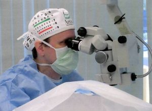
Age-Related Macular Degeneration (AMD): Early Detection and Treatment Options
Macular degeneration is one of the most common causes of vision loss in adults over age 55. What Is AMD? ...
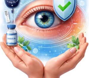
How Diabetes Affects Your Vision: Protecting Your Eyes Long-Term
Diabetes affects the entire body — and the eyes are one of the most vulnerable organs. Many diabetic patients develop ...
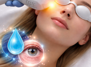
IPL Therapy for Dry Eye: A Modern Treatment for Meibomian Gland Dysfunction
IPL (Intense Pulsed Light) therapy is one of the most effective modern treatments for patients with chronic dry eye caused ...

Why Are My Eyes Always Dry? 7 Common Causes of Dry Eye Disease
If you find yourself constantly reaching for eye drops, you’re not alone. Chronic dry eye affects millions of Americans and ...
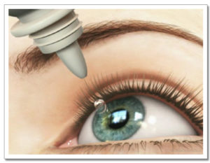
Chronic Dry Eye Syndrome: Causes, Symptoms, and Advanced Treatment Options
Dry eye disease is one of the most common reasons patients visit an ophthalmologist. Many patients think dry eye is ...